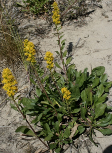
65 Spikelike Goldenrod
Names
Common name – Spikelike Goldenrod
Scientific name – Solidago spathulate
Traditional Indigenous Uses
Its roots and leaves could be chewed to ease the pain of a toothache or pressed against the cheek to bring comfort when the gums were sore. When the throat ached or became raw, the fresh leaves were chewed to soothe the pain and ease the swelling. The plant was used to stop bleeding and to heal wounds. The leaves were crushed into a poultice and placed on cuts, burns, or sores to draw out infection and help the skin close.
For bleeding on the inside, the people made a strong tea from the flowers and aerial parts, sometimes mixing it with the bark of the river birch to strengthen its healing power. The same tea was used for problems of kidneys and bladder, helping the body release water and wash away stones or infections. When the stomach was in pain, or digestion was troubled, the Goldenrod’s leaves and flowers were brewed into a gentle drink to calm the body and restore balance.
That the plant’s medicine also helped those who struggled to breathe. Steam and tea cleared the chest and loosened coughs. When sickness or fever came, the people would drink warm tea to cool their body and bring rest. The Goldenrod was also used to wash wounds and infections, for it cleansed the skin as well as the blood. Whether used inside or out, it reduced swelling and brought down inflammation.
Biochemical Basis for Medicinal Properties
Major Bioactive Compounds in Goldenrod
- Flavonoids (Primary Active Constituents)
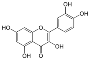
(i) Quercetin (C₁₅H₁₀O₇)
- Molecular structure: Pentahydroxyflavone with three aromatic rings (A, B, C rings)
- Contains 5 hydroxyl (-OH) groups at positions 3, 5, 7, 3′, and 4′
- Molecular weight: 302.24 g/mol
- These hydroxyl groups act as hydrogen donors and electron donors, which is crucial for antioxidant activity
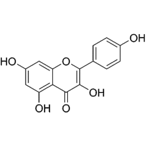
(ii) Kaempferol (C₁₅H₁₀O₆)
- Similar structure to quercetin but with 4 hydroxyl groups
- Molecular weight: 286.24 g/mol
- Lacks one hydroxyl group compared to quercetin (at position 3′)
(iii) Rutin (Quercetin-3-rutinoside, C₂₇H₃₀O₁₆)
- Glycosylated form of quercetin with rutinose sugar attached
- Molecular weight: 610.52 g/mol
- Better water solubility than quercetin alone
- Phenolic Acids
(i) Chlorogenic Acid (C₁₆H₁₈O₉)
- Ester formed between caffeic acid and quinic acid
- Strong antioxidant and anti-inflammatory properties
(ii) Caffeic Acid (C₉H₈O₄)
- Hydroxycinnamic acid derivative
- Contains catechol group (two adjacent hydroxyl groups on benzene ring)
- Saponins
Virgaureasaponins and Solidagosaponins
- Oleanane-type triterpene saponins
- Structure: 30-carbon triterpene backbone + oligosaccharide chains
- Amphipathic molecules (hydrophobic aglycone + hydrophilic sugar chains)
- Essential Oils and Terpenes
(i) Monoterpenes and Sesquiterpenes
- α-pinene, β-pinene, limonene, myrcene
- Volatile aromatic compounds
(ii) Diterpenes
- Labdane and clerodane types
- Anti-inflammatory activities
- Polysaccharides
- Complex carbohydrate structures
- Immunomodulatory properties
Chemical Reactions and Biochemical Mechanisms
- Antioxidant Mechanisms (Free Radical Scavenging)
Reaction 1: Hydroxyl Radical Scavenging by Quercetin
Quercetin-OH + •OH → Quercetin-O• + H₂O
(Flavonoid with hydroxyl) + (hydroxyl radical) → (flavonoid radical) + water
The hydroxyl groups on quercetin donate hydrogen atoms to neutralize highly reactive hydroxyl radicals (•OH), converting them to water. The quercetin molecule becomes a relatively stable phenoxyl radical (quercetin-O•) due to resonance stabilization across the aromatic rings.
Reaction 2: Superoxide Anion Scavenging
Quercetin-OH + O₂•⁻ + H⁺ → Quercetin-O• + H₂O₂
(Flavonoid) + (superoxide radical) + (proton) → (flavonoid radical) + hydrogen peroxide
Quercetin neutralizes superoxide radicals by electron donation. The resulting hydrogen peroxide is then metabolized by catalase.
Reaction 3: Metal Chelation
2 Quercetin + Fe³⁺ → [Quercetin₂-Fe³⁺] complex
The catechol groups (adjacent hydroxyl groups) on the B-ring and the 3-hydroxyl/4-carbonyl groups chelate transition metals like Fe³⁺ and Cu²⁺, preventing them from catalyzing Fenton reactions that produce damaging hydroxyl radicals.
2. Anti-inflammatory Mechanisms
Mechanism 1: Cyclooxygenase (COX) Inhibition
Flavonoids inhibit COX-1 and COX-2 enzymes by:
Arachidonic Acid + O₂ –[COX enzyme]–> Prostaglandin H₂ (PGH₂)
(INHIBITED by flavonoids)
PGH₂ → Prostaglandins (PGE₂, PGF₂α) + Thromboxanes
Mechanism: Flavonoids compete with arachidonic acid for the COX active site, reducing prostaglandin synthesis. Prostaglandins mediate pain, fever, and inflammation.
Mechanism 2: Lipoxygenase (LOX) Inhibition
Arachidonic Acid –[5-LOX enzyme]–> 5-HPETE → Leukotrienes (LTB₄, LTC₄)
(INHIBITED by quercetin)
Leukotrienes promote inflammation, bronchoconstriction, and immune cell recruitment. Quercetin inhibits 5-lipoxygenase, reducing leukotriene production.
Mechanism 3: NF-κB Pathway Inhibition
Stimulus (e.g., cytokines) → IκB kinase activation → IκB phosphorylation →
NF-κB release → Nuclear translocation → Pro-inflammatory gene transcription
(Quercetin inhibits IκB kinase and prevents NF-κB activation)
This reduces production of inflammatory cytokines (TNF-α, IL-1β, IL-6), adhesion molecules, and inducible nitric oxide synthase (iNOS).
3. Antimicrobial Mechanisms
Mechanism 1: Bacterial Membrane Disruption
Saponins interact with bacterial membrane sterols and phospholipids:
Saponin + Membrane cholesterol → Pore formation → Cell lysis
Hydrophobic triterpene inserts into lipid bilayer
Hydrophilic sugars create hydrophilic pores
Result: Loss of membrane integrity, ion leakage, cell death
Mechanism 2: Protein Denaturation
Phenolic compounds denature bacterial proteins:
Phenolic-OH + Protein-NH₂/SH groups → Hydrogen bonding/cross-linking →
Protein precipitation and inactivation
Mechanism 3: DNA Intercalation
Planar flavonoid structures can intercalate between DNA base pairs, interfering with replication:
Flavonoid molecules insert between purine-pyrimidine base pairs →
Distortion of DNA helix → Inhibition of DNA replication and transcription
4. Diuretic Mechanisms
Mechanism 1: Inhibition of Na⁺/K⁺-ATPase in Kidney Tubules
Flavonoids inhibit: 3Na⁺(inside) + ATP → 3Na⁺(outside) + 2K⁺(inside) + ADP + Pi
This reduces sodium reabsorption, increasing sodium and water excretion.
Mechanism 2: Increased Glomerular Filtration
Flavonoids cause vasodilation of renal arterioles through nitric oxide (NO) release:
L-Arginine –[eNOS]–> Nitric Oxide (NO) + L-Citrulline
(enhanced by flavonoids)
NO → Activation of guanylate cyclase → Increased cGMP → Smooth muscle relaxation →
Vasodilation → Increased renal blood flow → Enhanced glomerular filtration
5. Wound Healing Mechanisms
Mechanism 1: Collagen Synthesis Stimulation
Proline + α-ketoglutarate + O₂ + Ascorbate –[Prolyl hydroxylase]–> 4-Hydroxyproline (essential for collagen stability)
(Flavonoids enhance this process and protect ascorbate from oxidation)
Mechanism 2: Fibroblast Proliferation
Quercetin activates growth factor signaling:
Growth factors (PDGF, TGF-β) → Receptor tyrosine kinases → MAPK/ERK pathway activation → Fibroblast proliferation and migration
Mechanism 3: Anti-proteolytic Activity
Saponins and flavonoids inhibit matrix metalloproteinases (MMPs):
MMPs (collagenase, elastase) break down ECM proteins
Flavonoids inhibit MMP activity → Preserves extracellular matrix integrity → Supports tissue regeneration
6. Analgesic (Pain Relief) Mechanisms
Mechanism 1: Modulation of Opioid Receptors
Some triterpene saponins interact with μ-opioid receptors:
Saponin binds to μ-opioid receptor → G-protein activation → Decreased cAMP → Reduced Ca²⁺ influx → Decreased neurotransmitter release → Reduced pain signal transmission
Mechanism 2: Voltage-Gated Ion Channel Modulation
Flavonoids can block voltage-gated sodium channels (Nav) in nociceptors:
Membrane depolarization → Nav channels open → Na⁺ influx → Action potential
(BLOCKED by flavonoids)
Result: Reduced neuronal excitability and pain signal propagation
Summary of Structure-Activity Relationships
- Multiple Hydroxyl Groups: The phenolic -OH groups act as hydrogen donors for antioxidant activity and form hydrogen bonds with biological targets.
- Planar Aromatic Systems: Allow π-π stacking interactions with proteins and DNA, and provide resonance stabilization of radical intermediates.
- Amphipathic Nature (Saponins): Enables interaction with both lipid membranes and aqueous environments, crucial for antimicrobial activity.
- Chelation Sites: Catechol groups and keto-enol systems chelate metal ions, preventing oxidative damage.
- Lipophilicity: Allows compounds to cross cell membranes and reach intracellular targets.
References
Due to the limited specific published research on Solidago spicata Indigenous uses, these references primarily cover broader Solidago species ethnobotany and phytochemistry:
1) Elders and Community members of the Cayoose Creek Band of Sekw’el’was
2) Moerman, D. E. (1998). Native American ethnobotany. Timber Press.
3) Foster, S., & Duke, J. A. (2000). A field guide to medicinal plants and herbs of eastern and central North America (2nd ed.). Houghton Mifflin Company.
4) Apáti, P., Szentmihályi, K., Kristó, S. T., Papp, I., Vinkler, P., Szoke, É., & Kéry, Á. (2003). Herbal remedies of Solidago: Correlation of phytochemical characteristics and antioxidative properties. Journal of Pharmaceutical and Biomedical Analysis, 32(4–5), 1045–1053. https://doi.org/10.1016/S0731-7085(03)00213-0
5) Deng, Y., Zhao, Y., Padilla-Zakour, O., & Yang, G. (2015). Polyphenols, antioxidant and antimicrobial activities of leaf and bark extracts of Solidago canadensis Industrial Crops and Products, 74, 803–809. https://doi.org/10.1016/j.indcrop.2015.05.046
6) Kalemba, D., Thiem, B., & Góra, J. (1990). Seasonal variation in the chemical composition of the essential oil of Solidago virgaurea from natural habitat. Journal of Essential Oil Research, 2(4), 205–208. https://doi.org/10.1080/10412905.1990.9697860
7) Thiem, B., & Goślińska, O. (2004). Antimicrobial activity of Solidago virgaurea from in vitro cultures. Fitoterapia, 75(7–8), 668–673. https://doi.org/10.1016/j.fitote.2004.05.003
8) Reznicek, G., Jurenitsch, J., Plasun, M., Korhammer, S., Haslinger, E., Hiller, K., & Kubelka, W. (1991). Four major saponins from Solidago canadensis. Phytochemistry, 30(5), 1629–1633. https://doi.org/10.1016/0031-9422(91)84277-R
9) Bader, G., Kulhanek, M., & Hiller, K. (1992). Saponins from the aerial parts of Solidago virgaurea. Planta Medica, 58(2), 149–153. https://doi.org/10.1055/s-2006-961404
